The Anatomy of the Spinal Cord
- Title
- The Anatomy of the Spinal Cord
- Medium
- Line work technique
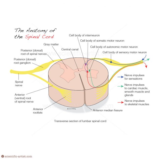
Illustration Description
This illustration of the transverse section of the lumbar spinal cord shows white matter in the periphery, gray matter inside, and a tiny central canal filled with CSF at its centre. Surrounding the canal is a single layer of cells, the ependymal layer. The spinal nerves extend form the spinal cord sending and receiving nerve impulses from and to the cardia, smooth and glands.
It was created using Adobe Illustrator and formed part of a series of card designs with an anatomical theme.