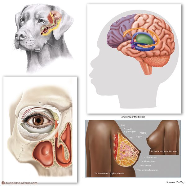
Human and Animal Anatomy Illustration

As a medical and scientific artist, my passion lies in studying and accurately illustrating bodily structures in a 2D format. This includes gross anatomy illustrations of organs and organ systems, as well as histology, which focuses on microscopic structures like cells, microbes, and organelles. I enjoy illustrating both human and animal anatomy, drawing inspiration from preserved specimens at museums, visits to the anatomy department at the Royal College of Veterinary Surgeons in London, and extensive reading. To see some animal anatomy examples you can view my gallery of illustrations.
Working as an anatomical artist requires extensive training in anatomy, which is crucial for accurately depicting the subject matter. Understanding both human and animal body’s inner workings—how ligaments attach to bones, the physiology of muscle fibres, and the subtleties of anatomical structures—is essential for creating precise illustrations. This deep knowledge ensures that the artwork serves its educational purpose effectively, providing accurate and detailed visuals for learning and teaching. To see some of my human anatomy artwork you can view my gallery of illustrations here.
During my career, I have had the privilege of attending surgery to create medical illustrations. This includes observing keyhole surgery for hernias, total knee replacements, hammer toe corrections, and small bowel surgeries. Over the past 15 years, I have collaborated closely with medical professionals, gaining in-depth knowledge through hands-on experience.